Researchers from UC San Francisco and Cedars-Sinai have developed a new way to prompt stem cells to form specific organs. It sets the stage for growing human organs from scratch – a long-time goal of regenerative medicine.
The research team showed that a few “organizer” cells can be programmed to coax other stem cells to form rudimentary, organ-like structures – including one that contracts like a beating heart and has a cavity resembling a heart ventricle.
“To build organs, we need to understand the rules behind their natural development,” said Wendell Lim, PhD, a professor of cellular and molecular pharmacology at UCSF. Lim is co-senior author of the paper, which appears Dec. 19 in Cell. “We wrote our own simple developmental programs from scratch, which we think could lead to more effective ways to repair and regenerate the body.”
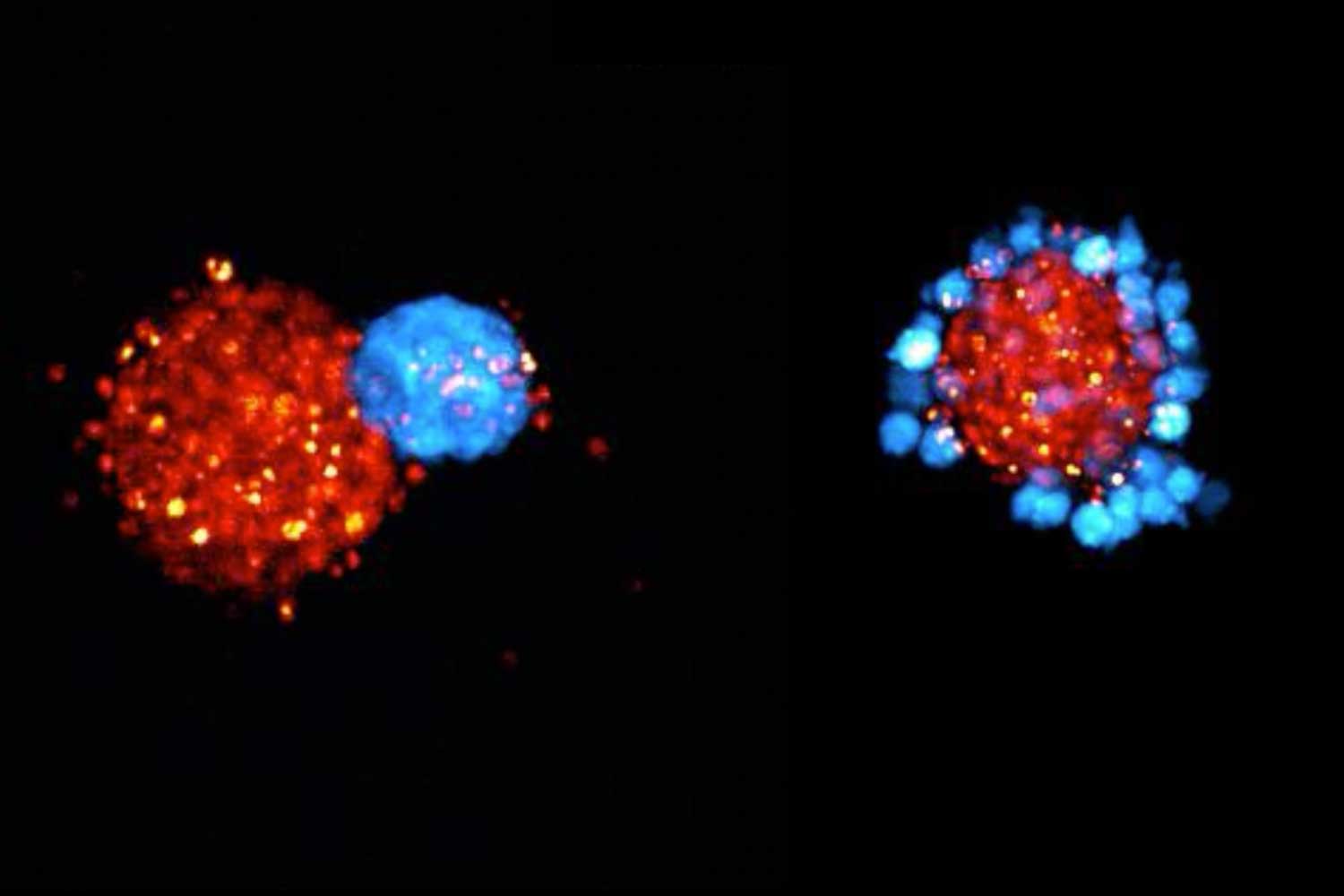
“Heart timelapse:” Organizer cells (blue), gathered as a node on top of a cluster of stem cells (outlined), instruct those stem cells to mature into different body tissues. The stem cells far from the node formed a small chamber, akin to a heart ventricle, and beat rhythmically, like the heart.
Credit: Toshi Yamada, Lim Lab.
Coaching cells through a developmental plan
Earlier attempts to grow organs in the lab relied on the ability of some cells, like those in the placenta, to instruct stem cells to begin to mature into more complex tissues. But these natural “organizer” cells can only implement a single growth blueprint, limiting their utility in regenerative medicine.
These engineered ‘organizer’ cells could someday enable us to repair and replace organs in the patients that need it.”
Lim and co-senior author Ophir Klein, MD, PhD, executive vice dean of Children’s Health at Cedars-Sinai and executive director of Cedars-Sinai Guerin Children’s, wanted more control over the process.
So, they engineered synthetic “organizer” cells to gather near stem cells and send them growth signals of their choosing.
They tested two arrangements of organizer cells around the stem cells: a node and a ring. With the node shape, stem cells close to the node received a stronger growth signal than cells that were further away. With a ring shape, stem cells inside the ring received a uniform amount of the growth signal.
How the organizer cells assembled around the stem cells, along with the signals they gave off, determined which genes the stem cells would express, taking them down different paths of maturation.
“By changing the placement of the organizer cells, and what signals they produced, we could finely tune how those signals spread through groups of stem cells,” said Klein, who is also an adjunct professor at UCSF. “In one case, we saw a gene expression response resembling early growth of the entire body. In another case, we got a response resembling just one region of the body.”

The first steps toward growing a heart
In another experiment in which the stem cells were instructed to become the cells of the central part of the body, they coalesced into roundish chambers that looked like the ventricles of the heart.
These structures contracted with a rhythmic beat and grew fine appendages resembling blood vessels. Beating cells have been grown from scratch in petri dishes before, but Lim said the well-defined chamber and vessel-like appendages represent a step forward for synthetic development.
“We really want to understand how the genome encodes a body plan and executes it,” said Lim, who directs the UCSF Cell Design Institute. “These engineered ‘organizer’ cells could someday enable us to repair and replace organs in the patients that need it.”
Authors: Other authors are Toshimichi Yamada, PhD, Coralie Trentesaux, PhD, Jonathan M. Brunger, PhD, Yini Xiao, PhD, Adam J. Stevens, PhD, Iain Martyn, PhD, Petr Kasparek, PhD, Neha P. Shroff, PhD, Angelica Aguilar, MS, Benoit G. Bruneau, PhD, all from UCSF; and Dario Boffelli, PhD, from Cedars-Sinai Medical Center.
Funding and disclosures: This work was funded in part by grants from the NIH (RO1 DK130969, RO1 HL114948, R35 DE026602) and the Nation Science Foundation (DBI-1548297), as well as support from the UCSF Cell Design Institute and the Valhalla Foundation. For all funding and disclosures, see the paper.