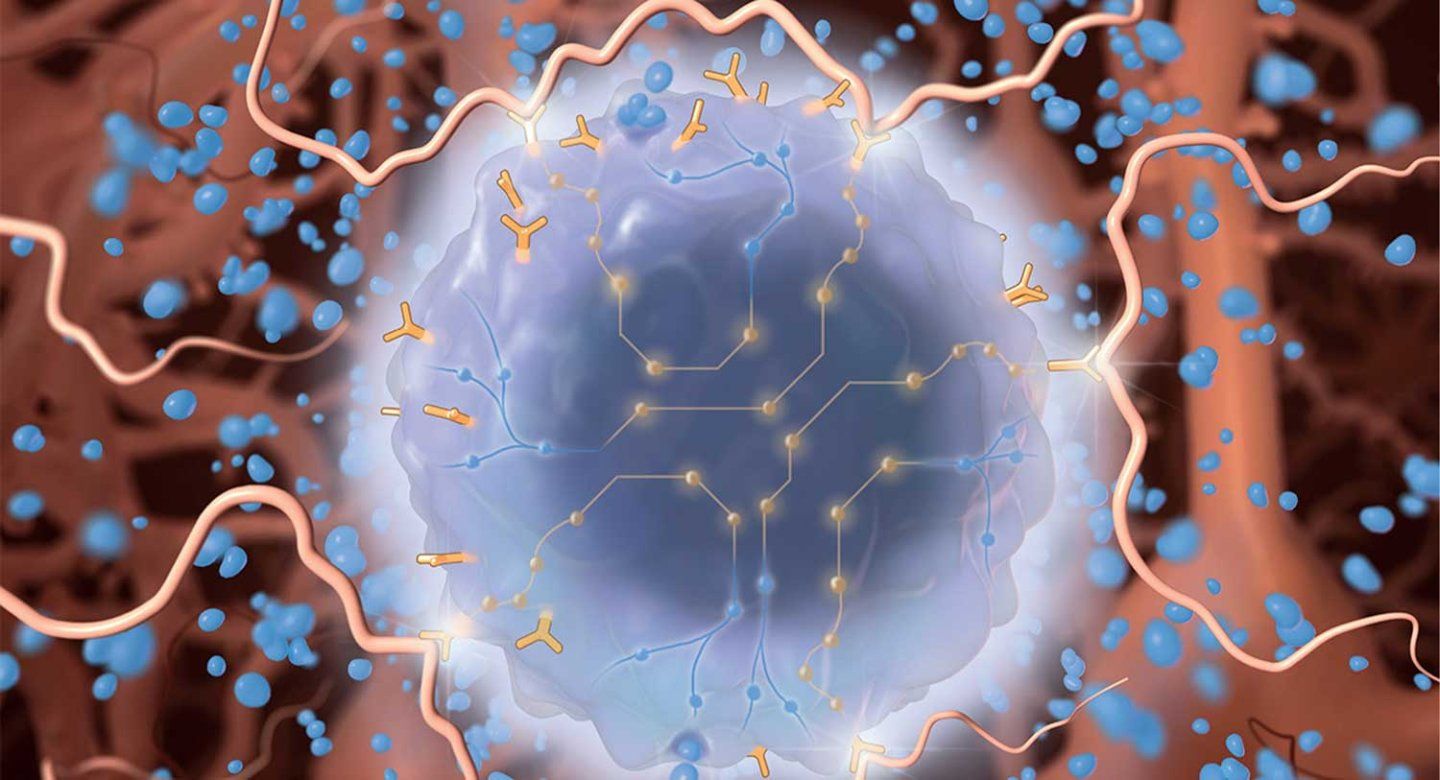
UCSF scientists have developed a “molecular GPS” to guide immune cells into the brain and kill tumors without harming healthy tissue.
It is the first living cell therapy that can navigate through the body to a specific organ, addressing what has been a major limitation of CAR-T cancer therapies until now. The technology worked in mice and the researchers expect it to be tested in a clinical trial next year.
Working in mice, the scientists showed how the immune cells could eliminate a deadly brain tumor called glioblastoma – and prevent recurrences. They also used the cells to tamp down inflammation in a mouse model of multiple sclerosis.
“Living cells, especially immune cells, are adapted to move around the body, sense where they are and find their targets,” said Wendell Lim, PhD, UCSF professor of cellular and molecular pharmacology and co-senior author of the paper, which appears in Science on Dec. 5.
Navigating to the source of disease
Nearly 300,000 patients are diagnosed with brain cancers each year in the U.S., and it is the leading cause of cancer mortality in children.
Because of their location, brain cancers are among the hardest cancers to treat. Surgery and chemotherapy are risky, and drugs can’t always get into the brain.
To get around these problems, the scientists developed a “molecular GPS” for immune cells that guided them with a “zip code” for the brain and a “street address” for the tumor.
They found the ideal molecular zip code in a protein called brevican, which helps to form the jelly-like structure of the brain, and only appears there. For the street address, they used two proteins that are found on most brain cancers.
The scientists programmed the immune cells to attack only if they first detected brevican and then detected one or the other of the brain cancer proteins.
When the scientists put the immune cells into the bloodstream, they easily navigated to the mouse’s brain and eliminated a growing tumor. Any immune cells that remain in the bloodstream stay dormant, sparing any tissues outside the brain that happen to have the same protein “address” from being attacked.
One hundred days later, the scientists introduced new tumor cells into the brain, and enough immune cells were left to find and kill them, a good indication that they may be able to prevent any remaining cancer cells from growing back.
Brain-sensing T cells (blue) turn green upon reaching the brain. These T cells, engineered in the Lim Lab, can be programmed to find a tissue like the brain (akin to a zip code), and then home in on the source of disease, like a tumor (akin to a street address). The approach successfully eliminated brain tumors in a mouse model.
“The brain-primed CAR-T cells were very, very effective at clearing glioblastoma in our mouse models, the most effective intervention we’ve seen yet in the lab,” said Milos Simic, PhD, the Valhalla Foundation Cell Design Fellow and co-first author of the paper. “It shows just how well the GPS ensured that they would only work in the brain. The same strategy even worked to clear brain metastases of breast cancer.”
In another experiment, the researchers used the brain GPS system to engineer cells that deliver anti-inflammatory molecules to the brain in a mouse model of multiple sclerosis. The engineered cells once again reached their target and made their delivery, and the inflammation faded.
The scientists hope this approach will soon be ready for patients with other debilitating nervous system diseases.
“Glioblastoma is one of the deadliest cancers, and this approach is poised to give patients a fighting chance,” said Hideho Okada, MD, UCSF oncologist and co-senior author of the paper.
“Between cancer, brain metastases, immune disease and neurodegeneration, millions of patients could someday benefit from targeted brain therapies like the one we’ve developed.”
Authors: Other UCSF authors are co-first author Payal B. Watchmaker, PhD, Sasha Gupta, MD, Sharon A Sagan, Jason Duecker, Chanelle Shepherd, David Diebold, Psalm Pineo-Cavanaugh, Jeffrey Haeglin, Robert Zhu, Ben Ng, Wei Yu, MD, PhD, Yurie Tonai, Nishith R. Reddy, PhD, Stephen L. Hauser, MD, Michael R. Wilson, MD, and co-senior author, Scott S. Zamvil, MD, PhD. For all authors see the paper.
Funding: The work was supported in part by grants from the Weill Institute for Neurosciences; the National Institutes of Health, NCI & NIBIB U54CA244438, NINDS R35NS105068, and NCI P50CA097257; ARPA-H D24AC00084-00; Living Therapeutic Initiative at UCSF; the Valhalla Foundation; UCSF Cell Design Institute; and HDFCCC Laboratory for Cell Analysis Shared Resource Facility NIH NCI award P30CA082103. For all funding see the paper.
Disclosures: Several patents have been filed related to this work (this includes but not limited to, US APP # 63/464,497; 17/042,032; 17/040,476; 17/069,717; 15/831,194; 15/829,370; 15/583,658; 15/096,971; 15/543,220). For all disclosures see the paper.
