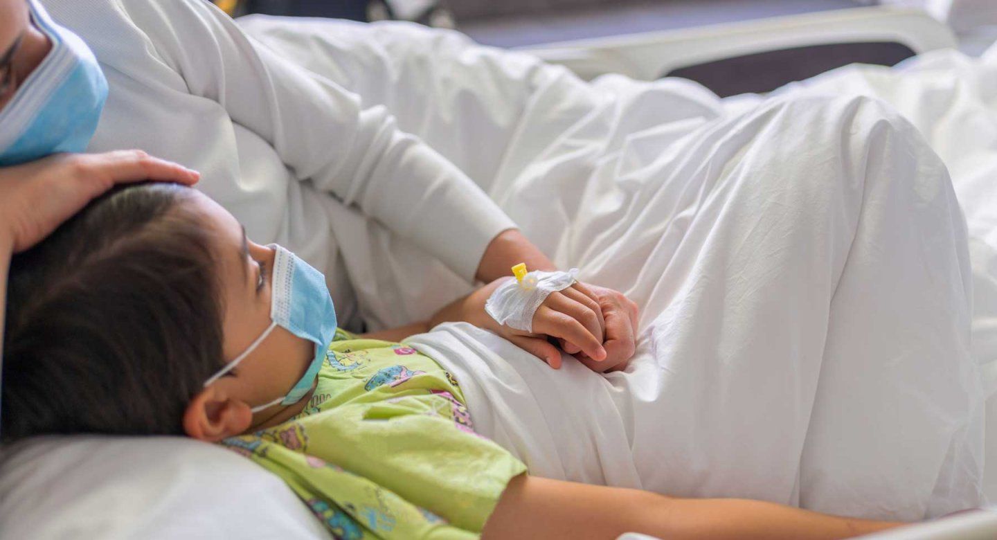
Early in the pandemic, some children fought off COVID with few, if any, symptoms, only to go into organ failure a few weeks later.
Most recovered after aggressive treatment, but their sudden illness, dubbed multisystem inflammatory syndrome in children (MIS-C), remained a mystery.
Now, a team of scientists from UC San Francisco, Chan Zuckerberg Biohub San Francisco, St. Jude Children’s Research Hospital and Boston Children’s Hospital has discovered what led to many of these cases, with a study that has implications for other autoimmune diseases.
The researchers found that the children’s immune systems had latched onto a part of the coronavirus that closely resembles a protein found in the heart, lungs, kidneys, brain, skin, eyes and GI tract, and launched a catastrophic attack on their own tissues.
The study, published Aug. 7 in Nature, offers one of the clearest connections yet linking viral infection and subsequent autoimmune disease.
“Thanks to our world-class team we’ve found an answer for how children get this mysterious disease,” said Aaron Bodansky, MD, a critical care fellow in UCSF’s Department of Pediatrics and lead author of the paper. “We hope this kind of approach can help break new ground in understanding similar diseases of immune dysregulation that have stumped us for decades, like multiple sclerosis or type 1 diabetes.”
COVID’s unexpected consequence in kids
As the novel coronavirus spread among millions of people, MIS-C cases mounted, affecting about 1 in 2,000 children under 18 with COVID.
Adrienne Randolph, MD, MSc, a critical care pediatrician at Boston Children’s Hospital and co-senior author of the paper, tracked these cases and collected patient samples through a national network of pediatric ICUs she had founded called Overcoming COVID-19.
“Every time COVID peaked in an area, about 30 days later, there’d be a peak of these kids presenting with what looked like septic shock in our network of ICUs, except they were negative for all kinds of infection,” Randolph said. “If we hadn’t intervened and supported them, they could have died.”
Reported MIS-C case ranges, on or before May 3, 2021
States with no cases reported: Maine, Vermont. States with 1 through 24 cases reported: Alaska, Delaware, Hawaii, Kansas, Montana, Nebraska, New Hampshire, New Mexico, North Dakota, Oklahoma, Rhode Island, South Dakota, West Virginia, Wyoming. States with 25 through 49 cases reported: Arkansas, Idaho, Iowa, Kentucky, Mississippi, Oregon, Washington, Washington D.C. States with 50 through 99 cases reported: Alabama, Colorado, Connecticut, Indiana, Minnesota, Missouri, Nevada, Ohio, Texas, Utah, Virginia, Wisconsin. States with 100 through 149 cases reported: Arizona, Georgia, Louisiana, Maryland, Massachusetts, Michigan, New Jersey, New York, North Carolina, Pennsylvania, South Carolina, Tennessee. States with 150 through 199 cases reported: Florida, Illinois. States with 300 or more cases reported: Califorinia.
Treating children with MIS-C at UCSF, Bodansky noticed that it resembled Kawasaki disease and other rare pediatric inflammatory diseases that he had long sought to better understand.
Co-senior author Mark Anderson, MD, PhD, who directs the Diabetes Center at UCSF and is a CZ Biohub SF Investigator, suggested Bodansky try a tool that he and Joe DeRisi, PhD, president of Chan Zuckerberg Biohub San Francisco, were using, called Phage Immunoprecipitation Sequencing (PhIP-Seq), to see if it could uncover the cause of MIS-C.
PhIP-Seq screens blood for autoantibodies, or antibodies that mistakenly attack the human body rather than an outside threat, such as a virus. DeRisi, corresponding author and professor of biophysics and biochemistry at UCSF, had already used PhIP-Seq to understand severe neurological symptoms, as well as the link between viral infections and multiple sclerosis.
“We thought, could there be some sort of trigger with the immune system that leads to MIS-C?” Anderson recalled. “Thanks to Overcoming COVID-19, which our pediatric ICU was part of, we realized there might be enough samples to do PhIP-Seq on kids that had MIS-C and compare their antibodies with kids that had gotten COVID but did not get MIS-C.”
A fingerprint of SARS-CoV-2 at the scene of disease
It opens the door to understanding why so many of these post-infectious, horribly inflammatory autoimmune events occur.”
With the approval of the Centers for Disease Control and Prevention (CDC), Randolph sent samples from 199 children with MIS-C to UCSF, along with 45 samples from children who had not developed MIS-C after COVID.
PhIP-Seq revealed that one third of the MIS-C cases had autoantibodies for an obscure human protein, called SNX8, which is present in certain immune cells that reside throughout the body. For some reason, the immune system was making antibodies targeting itself.
The scientists spent months hunting for a link between these autoantibodies and SARS-CoV-2. Then, late one night while looking at SNX8 on his computer screen with Bodansky, DeRisi had a flash of insight. What if the human protein resembled a portion of SARS-CoV-2’s N protein?
“We ran an analysis here on my computer – I did it myself – and it matched!” DeRisi recalled. “It was the same thing. It blew our minds."
Next, the team screened the MIS-C kids’ antibodies against proteins from the virus. The antibodies were targeting the very part of the N protein that matched SNX8. But antibodies from the healthy kids targeted other parts of the N protein that did not resemble a human protein.
The T-cell caper
The match between the autoantibodies, SNX8 and the N protein was a clear sign of an autoimmune overreaction.
But there was a hitch: antibodies can only go after threats that are floating in the blood. SNX8 is hidden inside of cells and would be invisible to them.
Anderson and Bodansky suspected that killer T cells, which screen the contents of human cells and destroy infected ones, were involved in this case of mistaken molecular identity.
Joe Sabatino, MD, PhD, UCSF professor of neurology and co-author of the paper, found that some T cells from the MIS-C patients seemed to target SNX8.
To prove that these T cells were truly attacking both targets, Paul Thomas, PhD, professor of host-microbe interactions at St. Jude, and his team carefully screened T cells from some of the MIS-C patients for matches with human SNX8 and the viral N protein.
Usually, doing an experiment like this is like trying to find a needle in a haystack. The body has so many different kinds of T cells that they need to be grown, or expanded, in the lab to find the rare culprit.
The MIS-C samples were different.
“Just taking these T-cells from patients directly, without any expansion, we found clusters of T cells that were specific for these two different target proteins,” said Thomas, who is a co-senior author of the paper. “It gives us an overwhelming amount of confidence that this response, associated with the autoantibody profile, is naturally elicited in a subset of these patients.”
Broad lessons for autoimmune disease
Most children who had MIS-C have fully recovered, and major ICUs now only see a few new cases each year, mostly in unvaccinated children.
By tracing the disease from virus to autoantibody to T cell, the findings point to a new way of investigating – and hopefully one day treating – autoimmune disease more generally.
“This paper is special because it actually finds the smoking gun – what made these kids so sick,” DeRisi said. “It opens the door to understanding why so many of these post-infectious, horribly inflammatory autoimmune events occur.”
Authors: Robert C. Mettleman (St. Jude) is co-first author. Other UCSF and/or Chan Zuckerberg Biohub SF authors are Sara E. Vazquez, Haleigh S. Miller, Andrew F. Kung, Elze Rackaityte, Colin R. Zamecnik, Jayant V. Rajan, Hannah Kortbawi, Caleigh Mandel-Brehm, Anthea Mitchell, Chung-Yu Wang, Aditi Saxena, Kelsey Zorn, David J. L. Yu, James Asaki, John V. Pluvinage, Michael R. Wilson, Natalie Cvijanovich, and Matt Zinter. For all authors, see the paper.
Funding and Disclosures: The work was supported by the National Institute of Child Health and Human Development (K12-HD000850), National Institute of Allergy and Infectious Diseases (R01AI154470, 5R01AI154470-03, 2R01AI136514-06, 3P01AI165077-01S1) and Chan Zuckerberg Biohub SF, among others. For all funding and disclosures, see the paper.
About UCSF: The University of California, San Francisco (UCSF) is exclusively focused on the health sciences and is dedicated to promoting health worldwide through advanced biomedical research, graduate-level education in the life sciences and health professions, and excellence in patient care. UCSF Health, which serves as UCSF’s primary academic medical center, includes top-ranked specialty hospitals and other clinical programs, and has affiliations throughout the Bay Area. UCSF School of Medicine also has a regional campus in Fresno. Learn more at ucsf.edu or see our Fact Sheet.
About the Chan Zuckerberg Biohub San Francisco: CZ Biohub San Francisco, a nonprofit biomedical research center founded in 2016, is part of the CZ Biohub Network, a group of nonprofit research institutes created and supported by the Chan Zuckerberg Initiative. CZ Biohub SF’s researchers, engineers, and data scientists, in collaboration with colleagues at our partner universities — Stanford University; the University of California, Berkeley; and the University of California, San Francisco — seek to understand the fundamental mechanisms underlying disease and develop new technologies that will lead to actionable diagnostics and effective therapies. Learn more at czbiohub.org/sf.